Atlas of Small Animal Ultrasonography
Wiley Blackwell (Verlag)
978-1-118-35998-3 (ISBN)
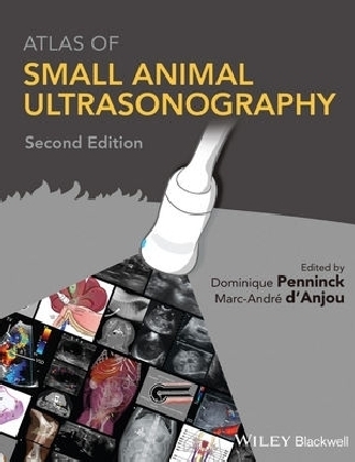
Atlas of Small Animal Ultrasonography is a comprehensive reference for ultrasound techniques and findings in small animal practice, with more than 2000 high-quality sonograms and illustrations of normal structures and disorders.
- Provides a comprehensive collection of more than 2000 high-quality images, including both normal and abnormal ultrasound features, as well as relevant complementary imaging modalities and histopathological images
- Covers both common and uncommon disorders in small animal patients
- Offers new chapters on practical physical concepts and artifacts and abdominal contrast sonography
- Includes access to a companion website with over 140 annotated video loops of the most important pathologies covered in each section of the book
Dominique Penninck, DVM, PhD, DACVR, DECVDI, is Professor of Diagnostic Imaging in the Department of Clinical Sciences, Cummings School of Veterinary Medicine, Tufts University.
Marc-André d'Anjou, DMV, DACVR, is Clinical Radiologist at Centre Vétérinaire Rive-Sud in the Montréal area, as well as at the Faculty of Veterinary Medicine of the Université de Montréal, where he was a professor for ten years.
Contributors
Preface
About the Companion Website
1. Practical Physical Concepts and Artifacts
Marc-André d'Anjou and Dominique Penninck
2. Eye and Orbit
Stefano Pizzirani, Dominique Penninck and Kathy Spaulding
3. Neck
Allison Zwingenberger and Olivier Taeymans
4. Thorax
Silke Hecht and Dominique Penninck
5. Heart
Donald Brown, Hugues Gaillot and Suzanne Cunningham
6. Liver
Marc-André d'Anjou and Dominique Penninck
7. Spleen
Silke Hecht andWilfried Mai
8. Gastrointestinal Tract
Dominique Penninck and Marc-André d'Anjou
9. Pancreas
Dominique Penninck and Marc-André d'Anjou
10. Kidneys and Ureters
Marc-André d'Anjou and Dominique Penninck
11. Bladder and Urethra
James Sutherland-Smith and Dominique Penninck
12. Adrenal Glands
Marc-André d'Anjou and Dominique Penninck
13. Female Reproductive Tract
Rachel Pollard and Silke Hecht
14. Male Reproductive Tract
Silke Hecht and Rachel Pollard
15. Abdominal Cavity, Lymph Nodes, and Great Vessels
Marc-André d'Anjou and Éric Norman Carmel
16. Clinical Applications of Contrast Ultrasound
Robert O'Brien and Gabriela Seiler
17. Musculoskeletal System
Marc-André d'Anjou and Laurent Blond
18. Spine and Peripheral Nerves
Judith Hudson and Marc-André d'Anjou
Index
| Zusatzinfo | illustrations |
|---|---|
| Verlagsort | New York |
| Sprache | englisch |
| Maße | 219 x 276 mm |
| Gewicht | 666 g |
| Einbandart | gebunden |
| Themenwelt | Medizin / Pharmazie ► Studium ► 1. Studienabschnitt (Vorklinik) |
| Veterinärmedizin ► Kleintier ► Bildgebende Verfahren | |
| Schlagworte | Hunde; Veterinärmedizin • Katzen; Veterinärmedizin • Kleintiere; Veterinärmedizin • Radiologie (Veterinärmedizin) |
| ISBN-10 | 1-118-35998-4 / 1118359984 |
| ISBN-13 | 978-1-118-35998-3 / 9781118359983 |
| Zustand | Neuware |
| Haben Sie eine Frage zum Produkt? |
aus dem Bereich


