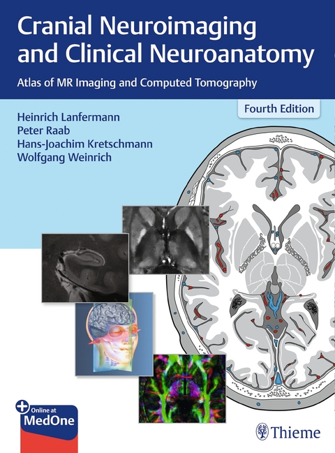
Cranial Neuroimaging and Clinical Neuroanatomy
Atlas of MR Imaging and Computed Tomography
Seiten
| Ausstattung: Softcover & Online Resource
2019
|
4th edition
Thieme (Verlag)
978-3-13-672604-4 (ISBN)
Thieme (Verlag)
978-3-13-672604-4 (ISBN)
Thieme's classic, indispensable guide to sectional imaging of the cranium
Now in a revised and expanded fourth edition, this exquisitely illustrated text/atlas by renowned experts, provides you with the cognitive tools to visualize and interpret CT and MR images of the cranium. In exacting detail, the normal structures of the brain, as seen in the three orthogonal planes (axial, sagittal, and coronal), are revealed with unparalleled accuracy, making the volume a highly useful aid in daily practice, for teaching, and to provide an anatomic baseline for research on the brain.
Beyond the clinical utility of the contents, the work is an aesthetic pleasure to behold, making learning and comprehension of complex material as simple and easy as possible.
Key Features:
New to the fourth edition:
»Cranial Neuroimaging and Clinical Neuroanatomy« is an essential reference guide for neuroradiologists and neurosurgeons (in training and in practice) and will also be welcomed by many neurologists.
Now in a revised and expanded fourth edition, this exquisitely illustrated text/atlas by renowned experts, provides you with the cognitive tools to visualize and interpret CT and MR images of the cranium. In exacting detail, the normal structures of the brain, as seen in the three orthogonal planes (axial, sagittal, and coronal), are revealed with unparalleled accuracy, making the volume a highly useful aid in daily practice, for teaching, and to provide an anatomic baseline for research on the brain.
Beyond the clinical utility of the contents, the work is an aesthetic pleasure to behold, making learning and comprehension of complex material as simple and easy as possible.
Key Features:
- Detailed brain anatomy shown in the three orthogonal planes; two-page spreads showing imaging studies keyed to the graphics using numbers that are consistent throughout
- Graphic representation of the major arterial and venous territories, and CNS spaces, supra- and infratentorial
- The most important neurofunctional systems revealed in multiplanar parallel sections, including detail on the potential sites of lesions and corresponding neurologic deficits
New to the fourth edition:
- All X-ray and CT-/MR images replaced with new high-resolution CT and MR images
- High resolution 3-Tesla MR images of the brainstem, 7-Tesla-images, fractional anisotropy (FA) maps as well as quantitative susceptibility maps (QSM)
- New material on temporal bone, brain maturation, neurofunctional systems
- Clinical context updated and expanded
»Cranial Neuroimaging and Clinical Neuroanatomy« is an essential reference guide for neuroradiologists and neurosurgeons (in training and in practice) and will also be welcomed by many neurologists.
lt;p>I Introduction
1 Introduction
2 Tomography and Landmarks
II Atlas
3 Coronal Sections
4 Sagittal Sections
5 Transverse Sections
6 Brainstem
III Topography of the Head and Neck
7 Topography of the Cranium, Intracranial Spaces, and Contained Structures
8 Facial Topography
9 Topography of the Head Neck Region
IV Nervous System Neurofunctional Systems and Neuroactive Substances
10 Neurofunctional Systems
11 Neurotransmitters and Neuromodulators
V Appendix
12 Specimens and Technique
13 Bibliography
| Erscheinungsdatum | 09.01.2019 |
|---|---|
| Zusatzinfo | 455 illustrations |
| Verlagsort | Stuttgart |
| Sprache | englisch |
| Maße | 230 x 310 mm |
| Gewicht | 2199 g |
| Einbandart | kartoniert |
| Themenwelt | Medizinische Fachgebiete ► Chirurgie ► Neurochirurgie |
| Medizin / Pharmazie ► Medizinische Fachgebiete ► Neurologie | |
| Medizinische Fachgebiete ► Radiologie / Bildgebende Verfahren ► Computertomographie | |
| Medizinische Fachgebiete ► Radiologie / Bildgebende Verfahren ► Kernspintomographie (MRT) | |
| Medizinische Fachgebiete ► Radiologie / Bildgebende Verfahren ► Neuroradiologie | |
| Schlagworte | Brain anatomy • brain imaging • Clinical Neuroanatomy • Computed tomography • cranial imaging • Cranial Neuroimaging • Cranium • CT • MRI • Neuroanatomy • neuroimaging • Neuroradiology • neurosurgery • Radiology |
| ISBN-10 | 3-13-672604-9 / 3136726049 |
| ISBN-13 | 978-3-13-672604-4 / 9783136726044 |
| Zustand | Neuware |
| Haben Sie eine Frage zum Produkt? |
Mehr entdecken
aus dem Bereich
aus dem Bereich
850 Fakten für die Zusatzbezeichnung
Buch | Softcover (2022)
Springer (Verlag)
49,99 €


