Rhoton's Atlas of Head, Neck, and Brain (eBook)
648 Seiten
Thieme Medical Publishers (Verlag)
978-1-60406-901-3 (ISBN)
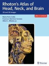
Masterful 2D and 3D head, neck, and brain dissections provide unsurpassed insights into head, neck, and brain anatomy
An internationally renowned and beloved author, educator, brain anatomist, and neurosurgeon, Professor Albert Rhoton has a special place in medical history. He was revered by students and colleagues and is regarded as one of the fathers of modern microscopic neurosurgery. A driving principle in his anatomy lab was the simple phrase, 'Every Second.' This was embraced in his philosophy that every second of every day, a patient's life was improved by a surgeon assisted by the anatomic knowledge his lab helped elucidate and distribute.
Rhoton's Atlas of Head, Neck, and Brain is the visually exquisite crowning achievement of Dr. Rhoton's brilliant career and unwavering dedication to the intertwined pursuits of surgical anatomy and neurosurgery. The atlas reflects the unparalleled contributions Dr. Rhoton made to the contemporary understanding of neurosurgical anatomy. Dr. Peris-Celda, with the collaboration of an impressive cadre of international multidisciplinary experts, worked closely under Dr. Rhoton's tutelage on this project. This book is the culmination of 5 years of work and experience gleaned from more than 40 years of surgical anatomy research and exquisite dissection techniques performed in Dr. Rhoton's laboratory.
Special Features
- Each anatomic dissection meticulously labeled with English and Latin descriptors for easy cross referencing with other resources.
- Multiple views of the most complex regions of the head, neck, and brain provide a deeper understanding of anatomy.
- More than 600 anatomical images systematically organized in four major sections: Osteology of the Head and Neck; Face and Neck; Ear, Nose, Pharynx, Larynx, and Orbit; and Neuroanatomy and Cranial Base.
- Superb 2D images presented in a large printed format to optimize the viewing experience.
- 3D digital images fully realize the beauty of the dissections and enhance the learning process.
- Specimens injected with colored silicone provide better visualization of arteries and veins.
Breathtakingly stunning, this atlas is certain to be a treasured reference for medical students, residents, and clinicians specializing in neurosurgery, facial plastic surgery, otolaryngology, maxillofacial surgery, and craniofacial surgery for many years to come.
Masterful 2D and 3D head, neck, and brain dissections provide unsurpassed insights into head, neck, and brain anatomyAn internationally renowned and beloved author, educator, brain anatomist, and neurosurgeon, Professor Albert Rhoton has a special place in medical history. He was revered by students and colleagues and is regarded as one of the fathers of modern microscopic neurosurgery. A driving principle in his anatomy lab was the simple phrase, "e;Every Second."e; This was embraced in his philosophy that every second of every day, a patient's life was improved by a surgeon assisted by the anatomic knowledge his lab helped elucidate and distribute.Rhoton's Atlas of Head, Neck, and Brain is the visually exquisite crowning achievement of Dr. Rhoton's brilliant career and unwavering dedication to the intertwined pursuits of surgical anatomy and neurosurgery. The atlas reflects the unparalleled contributions Dr. Rhoton made to the contemporary understanding of neurosurgical anatomy. Dr. Peris-Celda, with the collaboration of an impressive cadre of international multidisciplinary experts, worked closely under Dr. Rhoton's tutelage on this project. This book is the culmination of 5 years of work and experience gleaned from more than 40 years of surgical anatomy research and exquisite dissection techniques performed in Dr. Rhoton's laboratory.Special FeaturesEach anatomic dissection meticulously labeled with English and Latin descriptors for easy cross referencing with other resources.Multiple views of the most complex regions of the head, neck, and brain provide a deeper understanding of anatomy.More than 600 anatomical images systematically organized in four major sections: Osteology of the Head and Neck; Face and Neck; Ear, Nose, Pharynx, Larynx, and Orbit; and Neuroanatomy and Cranial Base.Superb 2D images presented in a large printed format to optimize the viewing experience.3D digital images fully realize the beauty of the dissections and enhance the learning process.Specimens injected with colored silicone provide better visualization of arteries and veins.Breathtakingly stunning, this atlas is certain to be a treasured reference for medical students, residents, and clinicians specializing in neurosurgery, facial plastic surgery, otolaryngology, maxillofacial surgery, and craniofacial surgery for many years to come.
| Erscheint lt. Verlag | 13.12.2017 |
|---|---|
| Zusatzinfo | Beilage: Online resource |
| Verlagsort | NEW YORK |
| Sprache | englisch |
| Themenwelt | Medizinische Fachgebiete ► Chirurgie ► Neurochirurgie |
| Medizin / Pharmazie ► Medizinische Fachgebiete ► HNO-Heilkunde | |
| Medizinische Fachgebiete ► Innere Medizin ► Pneumologie | |
| Medizin / Pharmazie ► Medizinische Fachgebiete ► Neurologie | |
| Studium ► 1. Studienabschnitt (Vorklinik) ► Anatomie / Neuroanatomie | |
| Schlagworte | 3D • brain • Dissection • English and Latin • Head • Microsurgery • neck • Peris-Celda • Rhoton • Surgical Anatomy |
| ISBN-10 | 1-60406-901-5 / 1604069015 |
| ISBN-13 | 978-1-60406-901-3 / 9781604069013 |
| Haben Sie eine Frage zum Produkt? |
Größe: 82,1 MB
DRM: Digitales Wasserzeichen
Dieses eBook enthält ein digitales Wasserzeichen und ist damit für Sie personalisiert. Bei einer missbräuchlichen Weitergabe des eBooks an Dritte ist eine Rückverfolgung an die Quelle möglich.
Dateiformat: PDF (Portable Document Format)
Mit einem festen Seitenlayout eignet sich die PDF besonders für Fachbücher mit Spalten, Tabellen und Abbildungen. Eine PDF kann auf fast allen Geräten angezeigt werden, ist aber für kleine Displays (Smartphone, eReader) nur eingeschränkt geeignet.
Systemvoraussetzungen:
PC/Mac: Mit einem PC oder Mac können Sie dieses eBook lesen. Sie benötigen dafür einen PDF-Viewer - z.B. den Adobe Reader oder Adobe Digital Editions.
eReader: Dieses eBook kann mit (fast) allen eBook-Readern gelesen werden. Mit dem amazon-Kindle ist es aber nicht kompatibel.
Smartphone/Tablet: Egal ob Apple oder Android, dieses eBook können Sie lesen. Sie benötigen dafür einen PDF-Viewer - z.B. die kostenlose Adobe Digital Editions-App.
Buying eBooks from abroad
For tax law reasons we can sell eBooks just within Germany and Switzerland. Regrettably we cannot fulfill eBook-orders from other countries.
aus dem Bereich