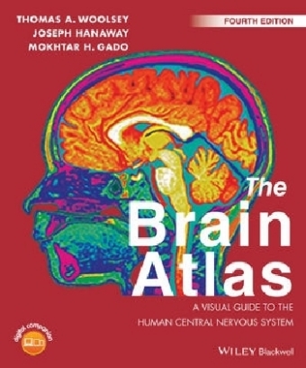
The Brain Atlas
John Wiley & Sons Inc (Verlag)
978-1-118-43877-0 (ISBN)
Dorian J. Pritchard is former lecturer in Human Genetics, University of Newcastle-upon-Tyne, UK and former Visiting Lecturer in Medical Genetics, International Medical University, Kuala Lumpur, Malaysia. Bruce Korf is Wayne H. and Sara Crews Finley Chair in Medical Genetics, Professor and Chair, Department of Genetics, and Director of the Heflin Center for Genomic Sciences at the University of Alabama at Birmingham, USA.
Preface
Acknowledgments
PART I
Introduction
Overview
The Nervous System
Cells
Gray Matter/White Matter
Connections
CNS/PNS
Constitution of the CNS
Principal Divisions of the Brain
Cranial Nerves and Spinal Segments
Cerebrospinal Fluid and Its Circulation
Cortical Areas
Using This Book
Terminology
Terms of Relation--The Special Case of the Human Head
Labels
Image Groups
Pathways
Navigation
Materials and Methods
Subjects
Brain Specimens
Radiological Imaging
References
PART II
The CNS and Its Blood Vessels
Brain
Cerebral Hemisphere and Brain Stem; Sulci and Gyri--Lateral Aspect
Cerebral Hemisphere and Brain Stem Arteries; Arteries of the Insula and Lateral Sulcus; Arterial Territories--Lateral Aspect
Cerebral Hemisphere and Brain Stem; Sulci and Gyri--Mesial Aspect
Cerebral Hemisphere and Brain Stem Arteries; Arterial Territories --Mesial Aspect
Cerebral Hemisphere and Brain Stem Arteries; by Conventional Angiography; by MRA--Lateral Projection
Dural Venous Sinuses and Folds (Diagrammatic); by Conventional Angiography; by MRV--Lateral Projection
Cerebral Hemispheres, Brain Stem and Arteries; by MRA--Anterior Aspect
Cerebral Hemisphere and Brain Stem Arteries and Veins by Conventional Angiography and Veins by MRV--Posteroanterior Projections
Cerebral Hemispheres and Brain Stem; Sulci and Gyri---Inferior Aspect
Cerebral Hemispheres and Brain Stem: Arteries and Cranial Nerves; Arterial Territories; Axial MRA--Inferior Aspect
Brain Stem
Brain Stem, Diencephalon, Basal Ganglia, and Cerebellum--Anterolateral Aspect
Brain Stem, Diencephalon, Basal Ganglia, and Cerebellum; Arteries and Cranial Nerves --Anterolateral Aspect
Brain Stem, Diencephalon, Basal Ganglia, and Cerebellum; Arterial Territories--Anterolateral Aspect
Brain Stem, Thalamus, and Striatum--Anterior Aspect
Brain Stem, Thalamus, and Striatum--Posterior Aspect
Brain Stem, Thalamus, and Striatum--Lateral Aspect
Cerebellum
Cerebellum--Superior Surface
Cerebellum--Inferior Surface
Spinal Cord
Arteries to Spinal Cord (Diagrammatic)
Segmental Arterial Supply of Spinal Cord (Diagrammatic)
Fiber Bundles
Principal Fiber Bundles in Cerebral Hemisphere and Brain Stem (Semi-Schematic)--Lateral and Mesial Aspects
Principal Fiber Bundles in Coronal, Axial, and Sagittal Brain Sections (Semi-Schematic)
PART III
Brain Slices
Coronal Sections
Coronal Section Through Rostral Wall of Lateral Ventricle with Vessel Territories
Coronal Section Through Anterior Limit of Putamen with MRI
Coronal Section Through Head of Caudate Nucleus and Putamen with MRI
Coronal Section Through Anterior Limit of Amygdala with Vessel TerritoriesCoronal Section Through Tuber Cinereum with MRI
Coronal Section Through Interventricular Foramen (Foramen of Monro) with Vessel Territories
Coronal Section Through Anterior Nucleus of Thalamus with MRI
Coronal Section Through Mamillothalamic Tract (Fasciculus) with Vessel Territories
Coronal Section Through Mamillary Bodies with MRI
Coronal Section Through Subthalamic Nucleus with Vessel Territories
Coronal Section Through Posterior Limit of Interpeduncular Fossa with MRI
Coronal Section Through Posterior Commissure with Vessel Territories
Coronal Section Through Commissure of Superior Colliculi with MRI
Coronal Section Through Quadrigeminal Plate with Vessel Territories
Coronal Section Through Fourth Ventricle (IV) with MRI
Coronal Section Through Posterior Limit of Hippocampus with Vessel Territories
Coronal Section Through Posterior Horns of Lateral Ventricles with MRI
Sagittal Sections
Sagittal Section Through Superior, Middle, and Inferior Temporal Gyri with Vessel Territories
Sagittal Section Through Insula with MRI
Sagittal Section Through Claustrum and Lateral Putamen with Vessel Territories
Sagittal Section Through Lateral Putamen with MRI
Sagittal Section Through Termination of Optic Tract with MRI
Sagittal Section Through Pulvinar with Vessel Territories
Sagittal Section Through Ambient Cistern with MRI
Sagittal Section Through Olfactory Tract with Vessel Territories
Sagittal Section Through Inferior Cerebellar Peduncle (Restiform Body) with Vessel Territories
Sagittal Section Through Superior Cerebellar Peduncle (Brachium Conjunctivum) with MRI
Sagittal Section Through Red Nucleus with Vessel Territories
Sagittal Section Through Cerebral Aqueduct (Aqueduct of Sylvius) with MRI
Axial Sections
Axial Section Through Superior Caudate Nucleus with MRI
Axial Section Through Inferior Corpus Callosum with Vessel Territories
Axial Section Through Superior Putamen with MRI
Axial Section Through Putamen with Vessel Territories
Axial Section Through Frontoparietal Opercula with MRI
Axial Section Through Midlevel Diencephalon with Vessel Territories
Axial Section Through Anterior Commissure with MRI
Axial Section Through Habenular Commissure with Vessel Territories
Axial Section Through Superior Colliculi with MRI
Axial Section Through Anterior Perforated Substance with Vessel Territories
Axial Section Through Inferior Colliculi with MRI
PART IV
Histological Sections
Cerebellum
Horizontal Section Through
Fastigial Nucleus
Horizontal Section Through
Dentate Nucleus
Brain Stem
Transverse Section Through Superior Colliculus with Vessel Territories
Transverse Section Through Oculomotor Nucleus
Transverse Section Through Inferior Colliculus with Vessel Territories
Transverse Section Through Superior Pons and Isthmus
Transverse Section Through Middle Pons with Vessel Territories
Transverse Section Through Facial Genu with MRI
Transverse Section Through Vestibulocochlear Nerve Root with Vessel Territories
Transverse Section Through Glossopharyngeal Nerve Root with Vessel Territories
Transverse Section Through Fourth Ventricle with Vessel Territories
Transverse Section Through Hypoglossal Nucleus with MRI
Transverse Section Through Inferior Olive with Vessel Territories
Transverse Section Through Decussation of Pyramids
Spinal Cord
Transverse Section Through Upper Cervical Level with Vessel Territories
Transverse Section Through Cervical Enlargement with MRI
Transverse Section Through Thoracic Level with Vessel Territories
Transverse Section Through Lumbar Enlargement with Vessel Territories
Transverse Section Through Sacral Level
Basal Ganglia and Thalamus
Coronal Section Through Nucleus Accumbens
Coronal Section Through Optic Chiasm
Coronal Section Through Anterior Commisure
Coronal Section Through Anterior Thalamic Tubercle
Coronal Section Through Mamillothalamic Tract
Coronal Section Through H Fields of Forel
Coronal Section Through Dorsal Lateral Geniculate Nucleus
Coronal Section Through Pulvinar
Hypothalamus
Coronal Section Through Optic Chiasm; Coronal Section Through Pituitary Stalk
Coronal Section Through Interthalamic Adhesion; Coronal Section Through Mamillary Bodies
Basal Forebrain
Coronal Section Through Olfactory Trigone and Nucleus Basalis
Hippocampus
Coronal Section Through Body Of Hippocampus
PART V
Pathways
Brain Stem
General Organization of Spinal Cord Gray Matter
General Organization of Cranial Nerve Gray Matter
Sensory Cranial Nerves and Nuclei
Motor Cranial Nerves and Nuclei
Organization of Cranial Nerve Nuclei into Columns--Posterior Aspect
Organization of Cranial Nerve Nuclei into Columns--Anterior Aspect
Thalamus
Hypothalamus
Sensory Systems
Touch and Position Sense Pathways: Posterior (Dorsal) Column/Medial Lemniscus and Trigeminal Main Sensory Nucleus
Touch Pathways: Anterior and Lateral Spinothalamic Tracts and Trigeminal Spinal Nucleus
Pain Pathways
Touch Pathways: Head and Face
Taste Pathways
Visual Pathways
Olfactory Pathways
Auditory Pathways R
Sensory/Motor Systems
Vestibular Pathways
Motor Systems
Corticospinal (Pyramidal) and Corticobulbar Pathways
Rubrospinal and Tectospinal Pathways
Reticulospinal Pathways
Cerebellum
Cerebellar Pathways: Somatic Afferents
Cerebellar Pathways: Afferents
(Non-Somatic)
Cerebellar Pathways: Efferents
Basal Ganglia
Basal Ganglia Pathways
Hippocampus
Hippocampal Pathways: Afferents
Hippocampal Pathways: Efferents
Amygdala
Amygdalar Pathways: Afferents
Amygdalar Pathways: Efferents
Hypothalamus
Hypothalamic Pathways: Afferents
Hypothalamic Pathways: Efferents
Arousal and Sleep
Arousal and Sleep Pathways
Hunger and Feeding
Circumventricular Organs
Autonomic Systems
Autonomic Pathways: Afferents
Autonomic Pathways: Sympathetic Efferents
Autonomic Pathways: Parasympathetic Efferents
Modulatory Systems
Cholinergic and Dopaminergic Pathways
Noradrenergic and Serotoninergic Pathways
Index
| Erscheint lt. Verlag | 17.4.2017 |
|---|---|
| Zusatzinfo | illustrations |
| Verlagsort | New York |
| Sprache | englisch |
| Maße | 231 x 275 mm |
| Gewicht | 804 g |
| Einbandart | kartoniert |
| Themenwelt | Geisteswissenschaften ► Psychologie |
| Medizin / Pharmazie ► Medizinische Fachgebiete ► Neurologie | |
| Studium ► 1. Studienabschnitt (Vorklinik) ► Anatomie / Neuroanatomie | |
| Studium ► 1. Studienabschnitt (Vorklinik) ► Physiologie | |
| Naturwissenschaften ► Biologie ► Humanbiologie | |
| Schlagworte | Neuroanatomie |
| ISBN-10 | 1-118-43877-9 / 1118438779 |
| ISBN-13 | 978-1-118-43877-0 / 9781118438770 |
| Zustand | Neuware |
| Haben Sie eine Frage zum Produkt? |
aus dem Bereich


