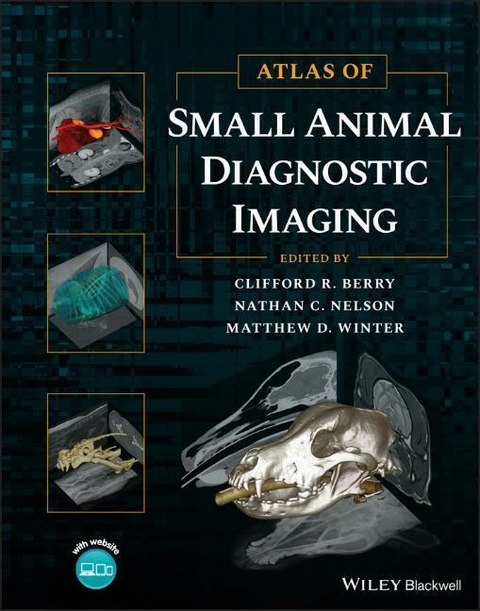
Atlas of Small Animal Diagnostic Imaging
John Wiley & Sons Inc (Verlag)
978-1-118-96440-8 (ISBN)
»Atlas of Small Animal Diagnostic Imaging« provides a comprehensive, multimodality atlas of small animal diagnostic imaging, with high-quality images depicting radiography, scintigraphy, ultrasonography, computed tomography, and magnetic resonance imaging.
Taking a traditional body systems approach, the book offers an image-intensive resource to survey radiographs with some other imaging modalities being used to emphasize interpretation of survey radiographs. The Atlas offers clinically relevant information for small animal practitioners and students.
Each body structure is thoroughly covered and well-illustrated, with discussion of the strengths and weaknesses of each modality in various scenarios.
Edited by three experienced radiographers, »Atlas of Small Animal Diagnostic Imaging« contains information on:
- Basics of diagnostic imaging, physics of diagnostic imaging, CT and MRI physics, US physics, and nuclear medicine physics
- Musculoskeletal normal anatomic variants, developmental orthopedic disease, joint disease, fracture and fracture healing, aggressive bone disease, and head and spine imaging
- Thorax anatomy, variants, and interpretation paradigm, extrathoracic structures, pleural space, pulmonary parenchyma, and mediastinum
- Abdomen anatomy, variants, and interpretation paradigm, extra-abdominal and body wall, peritoneal and retroperitoneal, liver and biliary, and spleen
With its expansive coverage of the subject and hundreds of high-quality images to aid in efficient and seamless reader comprehension, Atlas of Small Animal Diagnostic Imaging is an invaluable and must-have resource for small animal practitioners, veterinary students, veterinary radiologists, and specialists in a number of areas.
Clifford R. Berry, DVM, DACVR (DI), is a Courtesy Professor of Diagnostic Imaging at the College of Veterinary Medicine, University of Florida in Gainesville, FL, USA and Assistant Clinical Professor of Diagnostic Imaging at the College of Veterinary Medicine, North Carolina State University in Raleigh, NC.
Nathan C. Nelson, DVM, MS, DACVR (DI and EDI), is a Professor of Diagnostic Imaging at the College of Veterinary Medicine, North Carolina State University in Raleigh, NC, USA.
Matthew D. Winter, DVM, DACVR (DI), is an Associate Professor of Diagnostic Imaging at the College of Veterinary Medicine, University of Florida in Gainesville, FL, USA and Medical Director of Telehealth Services, Vet-CT, in Orlando, Florida.
List of Contributors xxx
Acknowledgements xxx
Preface xxx
About the Companion Website xxx
Section I: Introduction and Physics
1. Introduction to Diagnostic Imaging Winter
2. Physics of Diagnostic Imaging Huynh, Huguet, Berry
3. CT and MRI Physics Huguet, Huynh, Berry
4. US Physics Huguet, Huynh, Berry
5. Nuclear Medicine Physics Huynh, Huguet, Berry
Section II: Musculoskeletal
6. Normal Anatomic Variants Nelson
7. Developmental Orthopedic Disease Huynh
8. Joint Disease Nelson
9. Fracture and Fracture Healing Nelson
10. Aggressive Bone Disease Porter, Nelson
11. Head Imaging Nelson
12. Spine Imaging Nelson
Section III: Thorax
13. Anatomy, Variants, and Interpretation Paradigm Huynh, Berry
14. Extrathoracic Structures Grosso, Berry
15. Pleural Space Huguet, Berry
16. Pulmonary Parenchyma Huguet, Berry
17. Mediastinum Hecht
18. Cardiovascular Huguet, Berry
19. Feline Thorax Larson, Berry
Section IV: Abdomen
20. Anatomy, Variants, and Interpretation Paradigm Huguet, Giglio, Berry
21. Extra-Abdominal and Body Wall Winter
22. Peritoneal and Retroperitoneal Winter
23. Liver and Biliary Winter
24. Spleen Oliveira
25. Gastrointestinal Tract Hoey
26. Pancreas Oliveira
27. Urogenital Huynh
28. Adrenal Glands and Lymph Nodes Huynh
Index
| Erscheinungsdatum | 28.04.2023 |
|---|---|
| Verlagsort | New York |
| Sprache | englisch |
| Gewicht | 666 g |
| Einbandart | gebunden |
| Themenwelt | Veterinärmedizin ► Kleintier ► Bildgebende Verfahren |
| ISBN-10 | 1-118-96440-3 / 1118964403 |
| ISBN-13 | 978-1-118-96440-8 / 9781118964408 |
| Zustand | Neuware |
| Haben Sie eine Frage zum Produkt? |
aus dem Bereich


