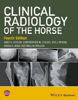
Clinical Radiology of the Horse
Wiley-Blackwell (an imprint of John Wiley & Sons Ltd) (Verlag)
978-1-4051-7108-3 (ISBN)
- Titel ist leider vergriffen;
keine Neuauflage - Artikel merken
Clinical Radiology of the Horse is the only book dedicated to the horse which provides a comprehensive overview of radiography and radiology of all areas of the horse. It provides a thorough guide both to the techniques used to obtain radiographs of the horse and to radiographic interpretation. With almost 600 superb annotated radiographs and more than 120 line diagrams, the book combines the best features of a high quality atlas and those of a detailed reference book. The normal radiographic anatomy of immature and mature horses is presented with normal variations, incidental findings and details of significant abnormalities. Remarks on clinical prognosis and treatment are also included. The emphasis throughout is on practical tips, common pitfalls, and the techniques used to obtain the best radiographs of specific areas and conditions.
Changes for the third edition: Significantly enlarged to include a chapter on digital radiography Includes descriptions of several new radiographic projections Many of the images have been replaced by digital images A wealth of new illustrations have been added Presents expanded information on processing and image quality Updated to include new information, knowledge gained from continued clinical experience and the most relevant references from recent literature CD included with the book presents all the radiographic images in electronic format Since publication of the Second Edition, there have been major advances in other imaging techniques, including scintigraphy, ultrasonography, computed tomography and magnetic resonance imaging. This third edition still focuses on radiography and radiology, but acknowledges the limitations of radiography in some circumstances. In these situations, reference is made to other imaging techniques which may be appropriate, along with suggestions for further reading.
Jan Butler Jan has specialized in equine radiography and has 30 years experience in this field. She currently works in private practice at the Willesley Equine Clinic in Gloucestershire, UK. Chris Colles BVetMed, PhD, Hon FWCF, MRCVS Chris is a senior partner in Avonvale Veterinary Practice, UK, specializing in equine orthopaedics. He is recognized by the Royal College of Veterinary Surgeons as a Specialist in Equine Orthopaedic Surgery. Sue Dyson MA, VetMB, PhD, DEO, FRCVS Sue specializes in lameness diagnosis and diagnostic imaging at the Centre for Equine Studies of the Animal Health Trust, Newmarket, UK. Sue is recognized as a specialist in Equine Orthopaedics by the Royal College of Veterinary Surgeons and holds the RCVS Diploma in Equine Orthopaedics. Svend Kold DVM, Dr Med Vet, CUEW, RFP, MRCVS Svend is a partner at the Willesley Equine Clinic, Gloucestershire, UK. He specializes in lameness and orthopaedic surgery and is recognized as a Specialist in Equine Orthopaedic Surgery by the Royal College of Veterinary Surgeons. Paul Poulos DVM, PhD, DipACVR Paul has his own consulting practice, Poulos Veterinary Imaging, based in Ukiah, California, having formerly held senior positions in academia at the University of Utrecht and the University of Florida. He has published widely on osteochondrosis, navicular disease and diseases of the fetlock.
Preface. 1. General Principles. 2. Computed and Digital Radiography. 3. Foot, Pastern and Fetlock. 4. The Metacarpal and Metatarsal Regions. 5. The Carpus and Antebrachium. 6. The Shoulder, Humerus, Elbow and Radius. 7. The Tarsus. 8. The Stifle and Tibia. 9. The Head. 10. The Spine. 11. The Pelvis and Femur. 12. The Thorax. 13. The Alimentary and Urinary Systems. 14. Miscellaneous Techniques. Appendix A: Fusion Times of Physes and Suture Lines. Appendix B: Exposure Guide, Image Quality and Film Processing Faults. Appendix C: Glossary. Index
| Erscheint lt. Verlag | 12.9.2008 |
|---|---|
| Zusatzinfo | Illustrations |
| Verlagsort | Chicester |
| Sprache | englisch |
| Maße | 227 x 274 mm |
| Gewicht | 3032 g |
| Themenwelt | Veterinärmedizin ► Klinische Fächer ► Bildgebende Verfahren |
| Veterinärmedizin ► Pferd ► Bildgebende Verfahren | |
| ISBN-10 | 1-4051-7108-1 / 1405171081 |
| ISBN-13 | 978-1-4051-7108-3 / 9781405171083 |
| Zustand | Neuware |
| Haben Sie eine Frage zum Produkt? |
aus dem Bereich